Paediatric Heart Surgery in India with the Best Paediatric Heart surgeons in India is what you would want if your little one is suffering from any heart ailment. MyMedOpinion affiliate Paediatric heart surgeons in India are among the top paediatric heart specialists and offer best in paediatric heart care. Affordable Pediatric Heart Surgery in India at Best Cardiac Hospitals with Top Pediatric Cardiac Surgeons in India with MyMedOpinion.
Sometimes heart surgery in children is required for repairing defects in the heart which a child might have born with (known as congenital heart defects) and heart diseases which he gets after birth, which require surgery. Pediatric Cardiac Surgery deals with operative procedures in the newborn and unborn children and youngsters suffering from cardiac dysfunctions, structural, functional and rhythm-related issues of the heart also.
In the normal heart, electrical impulses arise from an area of specialized cells called the sinus node, which is the heart's normal pacemaker. The sinus node is located in the right atrium, the upper right chamber of the heart . After leaving the sinus node, an impulse spreads across the upper heart chambers (right and left atria) and reaches the atrioventricular (AV) node located near the center of the heart between the atria and the lower chambers, or ventricles, and then to conduction fibers that spread across the ventricles, which are the heart's main pumping chambers.
As the impulse moves along the conduction fibers, cardiac muscle cells are stimulated and contract, producing a heartbeat.Pediatric Cardiac Surgery often deals with heart problems in children, the cause of congenital heart diseases which are common heart ailments among new born children and involves deficiencies like structural defects, congenital arrythmias and cardiomyopathies, which result in different kinds of abnormalities related to the heart. Congenital heart disease is a cause of improper growth of the heart or blood vessels before birth.

The most common cardiac defects in children are the following:
• Atrial Septal Defect
• Ventricular Septal Defect
• Fallot's Tetralogy
• Valvular defects
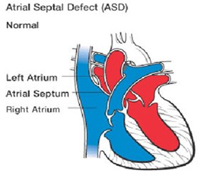
Atrial Septal Defect
An atrial septal defect (ASD) — sometimes referred to as a hole in the heart — is a type of congenital heart defect in which there is an abnormal opening in the dividing wall between the upper filling chambers of the heart (the atria). In most cases ASDs are diagnosed and treated successfully with few or no complications. When an atrial septal defect is present, blood flows through the hole primarily from the left atrium to the right atrium.
This shunting increases the blood volume in the right atrium which means more blood flows through the lungs than would normally. If left untreated, atrial septal defect may cause problems in adulthood. These problems may include pulmonary hypertension (which is high blood pressure in the lungs), congestive heart failure(weakening of the heart muscle), atrial arrhythmias (which are abnormal rhythms or beating of the heart) and an increased risk of stroke.
Ventricular Septal Defect - VSD have a hole in the wall of their heart between their right ventricle and left ventricle (the two lower chambers, where the blood leaves the heart). Normally, blood cannot pass between the ventricles. But when there is a hole between the sides of the heart, some oxygen-rich blood leaks from the left ventricle into the right ventricle and goes back to the lungs. The hole may be small and cause no symptoms, or it may be large and cause serious problems with blood flow. If the hole is large, too much blood will be pumped to the lungs, leading to congestive heart failure. Also, the heart will have to work harder to pump blood to the body. As a result of the extra work, the heart can get bigger.
There are two main techniques to correct the disorder:
Intra-Cardiac Technique - is an open heart surgery under general anesthesia , whereby the child is attached to an external heart-lung machine. This heart lung machine performs circulatory and respiratory function during surgery. The heart is directly accessed and the defect is sutured.
Trans- Catheter Technique - this is a minimally invasive technique through which surgical instruments are passed through the catheter inserted in the femoral artery. The catheter is slowly guided up towards the point of defect to close it.
Both the procedures are quite successful. Which one is best for your child can be ascertained by the paediatric cardiac surgeon in India after the examination of the child.
Fallot's Tetralogy
Fallot was a doctor who spotted this particular type of heart defect. Tetralogy means fourfold – there are four defects found together. These four problems are: 1. Pulmonary stenosis - Pulmonary means ‘of the lungs’. Stenosis means narrowing. Pulmonary stenosis is a narrowing at or below the pulmonary valve. 2. Ventricular septal defect -Ventricular means ‘of the ventricles’ – the wall between the right and left sides of the heart . 3. Over-riding aorta -The entrance to the aorta, which should only take red (oxygenated) blood around the body, lies over the VSD, allowing the right ventricle to pump some blue blood directly into it. 4. Right ventricular hypertrophy -The right ventricle becomes thickened as it forces blood into the narrowed pulmonary artery.
The heart surgeons perform two surgeries to correct Fallot of Tetralogy heart defect in the child Blalock- Taussig's operation is a palliative procedure performed in smaller infants to increase blood flow to lungs and to allow the child to grow big enough to withstand the corrective surgery. A connection is made between right subclavian artery and pulmonary artery to pass more oxygenated blood to the latter. This relieves the cyanosis to a great extent.
The total corrective surgery is performed in children within 2 years of age. VSD is closed with a patch and the narrowed pulmonary valve is opened. The outcome of surgery is favourable and most children lead a healthy life after the surgery with minimum restrictions
Double valve repair and replacement
Valves are openings between two chambers and also between a chamber and artery which allows unidirectional flow of blood. The patency of these valves is very important for normal flow of blood. The function of mitral valve (between left atrium and left ventricle) and the aortic valve (between left ventricle and aorta) is very important. If these valves get narrowed, the amount of blood passing from left atrium to left ventricle (mitral valve) or from left ventricle to aorta (aortic valve) is drastically reduced. In this case the heart will pump blood harder to push it through the narrowed valves. But the blood will tend to go upwards back into the pulmonary vein and finally to the lungs.
This will cause excess blood in the lungs leading to congestion. At the same time very less blood will come out into the aorta from the left ventricle which will result in reduced blood and oxygen supply to all the body parts.
Double valve repair and replacement procedure aims at correcting or replacing these both these damaged valves (aortic & mitral together) with new functional valves. This is done through the open heart surgery. The patient is put under general anaesthesia and connected to the heart lung machine. This machine takes over the pumping, circulatory, and respiratory functions of the heart and lung till the surgical procedure is going on. The valves are accessed by cutting open the rib cage and accessing the heart directly. The old valves are sliced from their attachments and new valves are put in their place.
The new valves may be obtained from a cadaveric donor, or an animal (pig) or it may be made of a nonreactive inert material. The patient needs to be given anticoagulants or immunosuppressive to clotting of blood or rejection of organ. The prognosis of this surgery is good. It is many a time a life-saving and life extending surgery.

Radio frequency catheter ablation is a procedure that is performed to correct a disturbance in heart rhythm (cardiac arrhythmia or irregular heartbeat).
It`s most often used to treat supraventricular tachyarrhythmias which are rapid, uncoordinated heartbeats starting in the heart`s upper chambers (atria) or middle region (AV node or the very beginning portion of the heart`s electrical system). In cardiology, RF ablation is used to correct:
• recurrent atrial flutter.
• atrial fibrillation (AF).
• supraventricular tachycardia (SVT).
• some types of ventricular arrhythmia.
About MyMedOpinion.com
MyMedOpinion affiliated Best hospitals in India provide an medical opinion from experienced surgeons and the treatment cost includes companion stay , surgeon fee, medicines and consumables, nursing care, patient's food and airport pick up & drop etc. etc. We offer free, no obligation assistance to international patients to find world class medical treatment in India. We offer support and services to facilitate the care you require. We can help you find the best hospital in India
Send us a Medical Report to Get FREE Medical Opinion from India's Top Doctors
MyMedOpinion.com is the hub of Surgeons and specialists for major diseases. We give you a chance to speak to our doctors and discuss your health issues directly.